I have always been interested with optics and vision. My path led me to a more technological approach of optics, aka photonics, but I still am fascinated by the process of vision. How the image is formed in the eye, how it is interpreted in the brain and so on. This led me to learn a few things that apparently are not so well known about our own eyes. One of these things is the subject of this post: Haidinger’s brush.
To start, it should come to no surprise to you that our eyes are sensitive to colours. We have 4 types of receptors in the retina, the part of the eye that is photosensitive. One of those type of receptor, the rod, is highly sensitive to light and responsible for night vision. The other 3 receptors are cones, and are sensitive to different portion of the light spectrum, either blue, green or red. Nothing amazing here.
The truly amazing thing that you might not know is that our eyes are also sensitive to the polarization of light. Polarization is a characteristic of light. To put it simply, polarization gives an idea of the direction of oscillation of the electric field. Polarization is independent of the wavelength. A linear polarization for a red light is the same as a linear polarization for a blue light and so on. There are a myriad of degree of polarization, ranging between a linear polarization (horizontal or vertical) to a circular polarization (circular right or circular left), with all sorts of elliptical polarisation in the middle. For this discussion, I’ll only talk about linear polarization.
How to see polarization? This trick requires to have access to a linear polarized source or light. As you might know, the light from a LCD monitor is linearly polarized. This is a characteristic of all the LCD screens that use crystals to bend the light polarization. And if you get a white background on that monitor or simply focus on some white space, you might be able to notice a small patch in the centre of your field of view that is not quite white…. It is actually a blue and yellow cross, although kind of hard to see. Typically, you won’t notice it if you don’t know it is there.
This cross is called Haidinger’s brush (or cross), discovered by an Austrian Physicist of the same name Wilhelm Karl von Haidinger [3] in 1844. You can see in figure 1 the type of result that you might be able to see, with the colour amplified of course… Haidinger’s brush is relatively small, taking approximately the size of your fovea, aka the spot in your field of view that you use to read. It has an angular size of a few degree only. The brush is two colour, a yellow line and a blue line perpendicular to the yellow line. And the orientation of the lines depend on the orientation of the polarization of the light.

One thing I need to add is that your eyes have that adaptability to colours (also called colour fatigue), meaning that if you look for the brush for too long, the brush might just fade away. To have more chances of seeing the brush, you can also rotate your head every second or two to change the orientation of the brush. I know, it will make you seem silly, but it can help. Don’t do that at work or in a public place, it will be hard to explain.
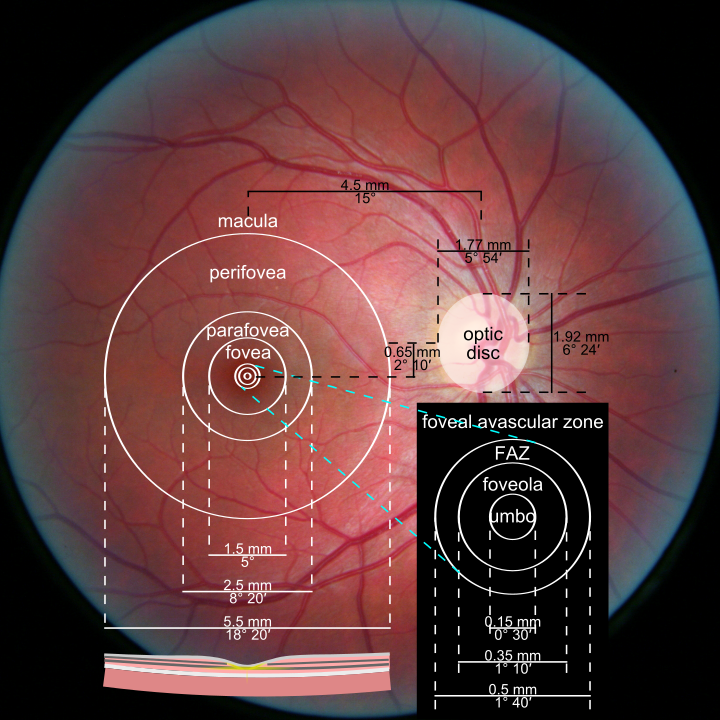
Why we see the Haidinger’s brush is still under debate by scientists [7], although there are some strong evidences that it is linked to the xanthophyll pigment [5]. This pigment is only present in our eye in the macula (see figure 2), the spot where we have the highest resolution, i.e. the portion of your field of view you are currently using to read those words. Actually, the full name of the macula is “macula lutea”, which means yellow spot. It turns out that the yellow comes from that specific pigment. Why don’t we see that yellow spot all the time in our vision? Because the brain ignores it, as simple as that.
The origin of the xanthophyll pigment and the reason why we would have it there is also interesting. It is a natural pigment that comes from vegetables, most notably carrots in the form of xanthophyll carotenoid. I guess that the saying that carrots are good for vision is not too far fetch after all. It has also been assumed that the reason why we have such pigment in our retina is to protect the sensitive cells behind from certain wavelengths, typically those in the blue region of the spectrum, which are quite energetic. This helps prevent damages to the retina, and is a marker for eye health typically. Some scientists think that seeing Haidinger’s brush could actually be used as a marker for eye health.
So we know that the xanthophyll pigment is yellow. It basically means that it is already absorbing blue light more than yellow to keep it simple. But this is not all: the xantophyll pigment is also dichroic. Dichroic means that the properties of the pigment (or any other material/molecule) depends with the wavelength and/or the polarization of the wavelength. The dichroism is actually going to accentuate this characteristics and make the blue polarized light absorbed much more efficiently than the yellow polarized light IF the polarisation of the light match the orientation of the molecule where the dichroic effect is maximum.
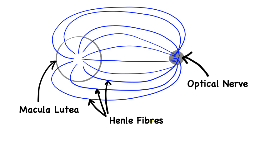
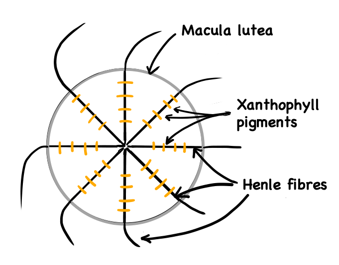
The dichroism alone is not enough to explain the appearance of the Brush. Most importantly is the fact that these pigments are arranged circularly inside the macula, perpendicular to the nerves coming from the optic nerve (the Henle fibers, see figures 3 and 4).
Because we take here a linear polarization, say horizontal, the absorption of the incoming white light from the screen is going to change depending on the orientation of the pigment. If the pigments are oriented in the right direction compared to the incoming polarization, that is the orientation where the dichroic effect is maximum, then the pigments are going to absorb more blue than normally, and the receptors in that region will detect a light slightly more yellow than usual. We have the opposite effect in the other region of the macula, where the polarization of the light does not match the orientation of the pigment. In that case, blue is abnormally going through more than usual, and the region appear more blue to the receptors. See figure 5. You have to remember that the eye response to white light, and more importantly the correction that the brain makes to make the colour adjustment, is “calibrated” to un-polarized light, which is also why the region at 50% appear white in the brush.
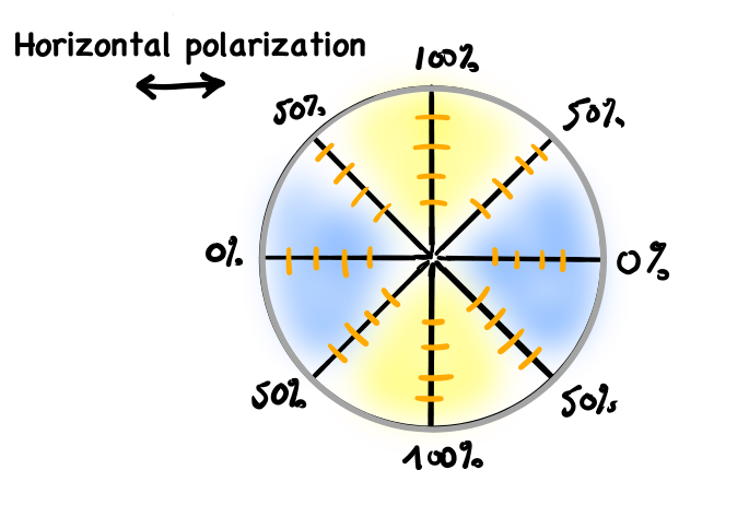
One can note that the yellow branch of the cross is always going to be perpendicular to the orientation of the linear polarization.
Once you master it on the computer screen, you also can try seeing the cross by looking in the sky, during a clear day, with a blue sky, when the sun is preferably low on the horizon and if you look straight up… I know, it is a lot of if, but again, it is to get that perfect condition where light will be polarized just enough to be able to see the pattern. Personally, I am not able to see it on the blue sky, but I see it on a white LCD screen.
Weirdly enough (I know, right?), this Haidinger’s brush is not a well known phenomenon, even by people who study optics and know about polarization. I think it is amazing the sort of hidden capacity, that everyone of us has, but we never actually notice them until someone is bored enough to just stare blankly in a white screen space and realize that there is a little spot of colour that appears in the middle of the field of view.
Sources
[1] http://www.polarization.com/haidinger/haidinger.html
[2] https://en.wikipedia.org/wiki/Haidinger%27s_brush
[3] https://en.wikipedia.org/wiki/Wilhelm_Karl_von_Haidinger
[4] https://en.wikipedia.org/wiki/Polarization_(waves)#Poincar%C3%A9_sphere
[5] https://en.wikipedia.org/wiki/Xanthophyll
[6] https://www.animations.physics.unsw.edu.au/jw/light/complementary-colours.htm
[7] https://www.nature.com/articles/s41598-019-56916-8
[8] https://en.wikipedia.org/wiki/Macula_of_retina
[9] https://en.wikipedia.org/wiki/Photoreceptor_cell#Humans
[10] http://hyperphysics.phy-astr.gsu.edu/hbase/phyopt/polabs.html
[11] https://www.sciencedirect.com/science/article/pii/S0042698908002289